PATTERNS OF HEPATIC MASS ENHANCEMENT
| ARTERIAL PHASE |
DLAYED PHASE IMAGES | DIAGNOSIS |
|---|---|---|
| Hyperenhancing | Hypoenhancing | HCC/hypervascular mets/enoma |
| Hypoenhancing | Hypoenhancing | Metastasis |
| Hyperenhancing | Isoenhancing | Focal Nodular Hyperplasia – FNH |
| Hyperenhancing | Hyperenhancing | Flash-filling hemangioma |
| Hypoenhancing | Hyperenhancing | Cholangio Carcinoma |
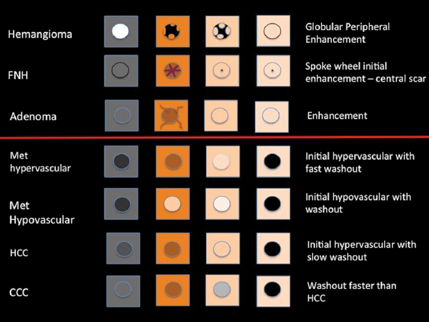
| Periph nodular | Centripetal fill in | Hemangioma – Classic |

Frequently Asked Questions
What are the common enhancement patterns of hepatic masses on CT and MRI?
Hepatic masses can show various enhancement patterns on imaging. Common patterns include hyperenhancing in the arterial phase and hypoenhancing in the delayed phase, typical of hepatocellular carcinoma (HCC) or hypervascular metastases. Focal Nodular Hyperplasia (FNH) often appears hyperenhancing in both phases, while classic hemangiomas show a peripheral nodular enhancement with centripetal fill-in. Understanding these patterns helps in diagnosing different types of liver lesions.
How does hepatocellular carcinoma appear on imaging studies?
Hepatocellular carcinoma (HCC) typically appears as a hyperenhancing mass during the arterial phase and becomes hypoenhancing during the delayed phase on CT and MRI. This pattern is due to the tumor's blood supply characteristics, which are different from normal liver tissue. Recognizing this enhancement pattern is crucial for the accurate diagnosis of HCC.
How can MRI help differentiate between types of liver lesions?
MRI is a powerful tool for differentiating liver lesions due to its ability to provide detailed contrast-enhanced images. For example, focal nodular hyperplasia (FNH) typically shows hyperenhancement in both arterial and delayed phases. In contrast, cholangiocarcinoma may appear hypoenhancing initially but becomes hyperenhancing in delayed images. These patterns assist radiologists and clinicians in making accurate diagnoses.
What is the significance of hypoenhancing hepatic lesions in imaging?
Hypoenhancing hepatic lesions, particularly when they remain hypoenhancing in both arterial and delayed phases, are often indicative of metastasis. This suggests that the lesion is not receiving as much blood supply as the surrounding liver tissue, a characteristic common in metastatic cancer. Identifying this pattern is important for diagnosing secondary liver cancers.
What distinguishes a hemangioma on liver imaging?
A classic hemangioma on liver imaging is characterized by a peripheral nodular enhancement pattern with centripetal fill-in over time on delayed phase images. This means that the enhancement starts at the edges and gradually fills towards the center. This pattern is distinct and helps differentiate hemangiomas from other liver lesions, as it reflects the lesion's blood vessel structure.