Table of Contents
What is Spondylosis?
Spondylosis (spinal osteoarthritis) is a degenerative disorder that can cause loss of normal spinal structure and function. Although aging is the primary cause, the location and rate of degeneration is individual. The degenerative process of spondylosis can affect the following regions of the spine: cervical (neck), thoracic (middle back), or lumbar (lower back).
Spine anatomy

The spine is made up of 31-34 bones (vertebral bodies) that are stacked on each other and are joined by ligaments. The spinal column can be divided into four different zones according to the characteristics of its vertebrae: Cervical spine (7 vertebrae), thoracic spine (12 vertebrae), lumbar spine (5 vertebrae) and sacro-coccygeal area (3 to 5 vertebrae).
An important feature of our spine is the physiological curves we have. If you look at the spine in profile you will find that it has a shape that resembles an “S”. These two curvatures are known as kyphosis and lordosis. Lordosis is that “sunken” curvature that we have in the neck (cervical region) and in the lower back (lumbar region); kyphosis is the curvature we have in the upper part of the back (thoracic region).
It is important to point out that contrary to popular belief, we should not be totally upright, these curvatures are natural and really necessary for the correct balance of forces in our spine.
Lumbar spine and its characteristics
The lumbar spine is made up of five vertebrae (L1, L2, L3, L4, and L5). These vertebrae are the largest in the entire spinal column and therefore can support most of the body weight.
The vertebrae are connected at the back by two joints called facet joints and in the space between them is the intervertebral disc. The intervertebral disc is very similar to a cushioning mattress and its function is to prevent injuries between each vertebral in addition to keeping them together.
Between the posterior elements of the vertebrae there are spaces or holes (foramina) through which the nerve roots from the spinal cord exit, which will then connect with each other to form the sciatic nerve, which extends to the legs on the back. back of each thigh to the feet.
Spondylosis often affects the following elements of the spine:
Intervertebral discs:
As people age, certain biochemical changes occur that affect the tissue found throughout the body. In the spine, the structure of the intervertebral discs (annulus fibrosus, lamella, nucleus pulposus) may be compromised. The annulus fibrosus (similar to a tire) is composed of 60 or more concentric bands of collagen fibers called lamellae. The nucleus pulposus is a gelatinous substance within the intervertebral disc enclosed by the annulus fibrosus. Collagen fibers form the core along with water and proteoglycans. The degenerative effects of age can weaken the structure of the annulus fibrosus, causing the “tire tread” to wear or tear. The water content of the core decreases with age, which affects its ability to rebound after compression (ie the quality of absorbing impacts). The structural alterations of the degeneration can decrease the height of the disc and increase the risk of a herniated disc.

Facet joints:
Each vertebral body has four facet joints that work like hinges. They are the movement joints of the spine that allow extension, flexion and rotation. Like other joints, the bony articulating surface is covered with cartilage. Cartilage is a special type of connective tissue that provides a low-friction, self-lubricating sliding surface. Facet joint degeneration leads to cartilage loss and osteophyte (ie, bone spur) formation. These changes can lead to hypertrophy or osteoarthritis, also known as degenerative joint disease.
Osteophytes in bones and ligaments (ie, bone spurs) can develop adjacent to the end plates, which can compromise the blood supply to the vertebra. Also, the end plates can become stiff due to sclerosis, a thickening or hardening of the bone beneath these plates. Ligaments are bands of fibrous tissue that connect the structures of the spine (ie, the vertebrae) and protect them against extremes of movement (ie, hyperextension). However, degenerative changes can cause the ligaments to lose some of their strength. The ligamentum flavus (a primary spinal ligament) may thicken or bulge posteriorly (back) toward the dura mater (a membrane of the spinal cord).
Symptoms of spondylosis and different levels of the spine
Cervical (neck):
The complexity of the cervical (neck) anatomy and its wide range of motion make this segment of the spine susceptible to disorders associated with degenerative change. Neck pain from spondylosis is common. The pain can spread to the shoulders and arms. When a bone spur (osteophyte) causes compression of a nerve root, the result can be weakness in the extremities (eg, the arms). In rare cases, bone spurs that form in the front of the cervical spine can cause difficulty swallowing (dysphagia).
Thoracic (middle back):
Forward bending and hyperextension often trigger the pain associated with degenerative disease. In the thoracic region of the spine, disc pain can be caused by flexion and facet pain by perextension.
Lumbar (lower back):
Spondylosis often affects the lumbar spine in people over 40 years of age. Morning pain and stiffness are common ailments. It is common for multiple levels to be involved (ie more than one vertebra). The lumbar spine supports most of the body weight. Therefore, when degenerative forces compromise structural integrity, activity may be accompanied by some symptoms, including pain. Movement stimulates pain fibers in the annulus fibrosus and facet joints. Sitting for long periods of time can cause pain and other symptoms due to pressure on the lumbar vertebrae. Repetitive movements such as lifting and bending (eg manual labor) can increase pain.
Risk factor’s for Spondylosis
Age is the most common risk factor for cervical spondylosis. The condition is extremely common in middle-aged and older patients.
Other factors that can increase the risk of developing cervical spondylosis and neck pain include the following:
- Genetics, family history of neck pain and spondylosis
- Smoking clearly linked to increased neck pain
- Occupation, jobs with a lot of repetitive neck movements or tasks at height
- depression or anxiety
- Previous neck injury or trauma
Symptoms of spondylosis
For most people, cervical spondylosis causes no symptoms. When symptoms do appear, they typically include neck pain and stiffness. This pain can be mild to severe. Sometimes it gets worse when looking up or down for a long time, or doing activities that keep the neck in the same position for a long time, such as driving or reading a book. The pain usually improves with rest or lying down.
Other symptoms may include the following:
- Headaches
- Grinding or pounding noise or sensation when turning the neck
- In some cases, cervical spondylosis causes narrowing of the space needed for the spinal cord and nerve roots. If this occurs, symptoms may include numbness and weakness in the arms, hands, and fingers.
- Difficulty walking, loss of balance, or weakness in the hands or legs
- Muscle spasms in the neck and shoulders
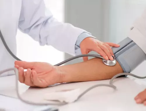
Medical examination for spondylosis
Physical exam
After discussing your medical history and general health, your doctor will perform a thorough exam of your neck, shoulders, arms, and often your legs. He will run a number of tests to see if there are any problems or changes in the following:
- Arm, hand and finger strength
- sensation of touch
- Reflexes
- Blood flow
- Flexibility in the neck and arms
- Andar (way of walking)
The doctor may also gently press on your neck and shoulders to check for trigger points (tender points) or swollen glands. They will also ask you questions to understand more about your symptoms and if you had any neck injuries. These questions may include the following:
- When did the pain start?
- When does the pain appear? Is it continuous or does it come and go?
- Do certain activities make the pain worse?
- Did you have pain before?
- Have you ever received treatment for pain?
- Do you have weakness or numbness in your arms or legs?
- Do you have difficulty with fine motor skills, such as writing or buttoning your shirt?
- Do you lose your balance or have other coordination problems?
- Have you ever been in an accident or had a neck injury?
Tests
The doctor may order diagnostic tests to confirm the diagnosis of cervical spondylosis. These tests may include the following:
X-rays. X-rays provide images of dense structures, such as bones. An x-ray will show the alignment of the bones in the neck. It can also show degenerative changes in the cervical spine, such as loss of disc height or the presence of bone spurs.
Magnetic resonance imaging (MRI) scans. MRI scans create better images of the body’s soft tissues, such as muscles, discs, nerves, and the spinal cord, than X-rays. An MRI can be used to determine if the symptoms are caused by soft tissue damage, such as herniated or bulging discs.
Computed tomography (CT) scans. A CT scan is more detailed than an X-ray and can help the doctor see the spinal canal and bone spurs better.
Myelogram. In this imaging procedure, a contrast dye is injected into the spinal canal to help the spinal cord and nerve roots show up more clearly. A CT scan is usually done after the myelogram while the contrast dye is still in the spinal canal.
Electromyography (EMG). Electromyography measures electrical impulses in muscles at rest and during contractions. Nerve conduction studies are often done with EMG to determine if a spinal nerve is working properly.
Other tests. In some cases, the doctor may order blood tests to determine if rheumatoid factor or other antibodies indicative of inflammatory arthritis are present.
Treatment options for spondylosis
non-surgical treatment
In most cases, treatment for cervical spondylosis is non-surgical. Non-surgical treatment options include the following:
Physiotherapy. Physical therapy is usually the first treatment your doctor will recommend. Some specific exercises can relieve pain, as well as strengthen and stretch weak or tight muscles. In some cases, physical therapy may include posture therapy or the use of traction to gently stretch the joints and muscles of the neck. The length of physical therapy programs varies, but they generally last 6-8 weeks. Typically, sessions are scheduled 2-3 times a week.
Medicines. During the first stage of treatment, your doctor may prescribe several medications to use together to treat pain and inflammation.
- Paracetamol. Mild pain is usually relieved with paracetamol.
- Nonsteroidal anti-inflammatory drugs (NSAIDs). Often prescribed with acetaminophen, NSAIDs , such as aspirin, ibuprofen, and naproxen, are considered first-line medications for neck pain. All relieve pain and inflammation, and may be prescribed for several weeks, depending on specific symptoms. Other types of pain medications may be considered if you have severe contraindications to NSAIDs or if your pain cannot be controlled.
- Oral corticosteroids. A short course of oral corticosteroids can help relieve pain and reduce inflammation.
- Muscle relaxants. Some medications, such as cyclobenzaprine or carisoprodol, can be used to treat painful muscle spasms.
Soft cervical collar. It is a padded ring that wraps around the neck and is fastened with Velcro. Your doctor may recommend that you wear a soft cervical collar to limit neck movement and allow your neck muscles to rest. A soft neck brace should only be worn for a short time, as prolonged use can decrease the strength of the neck muscles.
Cold, heat and other resources. Your doctor may recommend careful use of ice, heat, massage, and other local therapies to relieve symptoms.
Steroid injections. Many patients get short-term pain relief with steroid injections. The most common procedures for neck pain include the following:
- Cervical epidural block. In this procedure, steroid and anesthetic medications are injected into the space next to the lining of the spinal cord (epidural space). This procedure is typically used for neck or arm pain that may be due to a herniated cervical disc, also known as radiculopathy or pinched nerve.
- Cervical facet joint block. In this procedure, steroid and anesthetic medications are injected into the capsule of the facet joint. The facet joints are located at the back of the neck and provide stability and movement. These joints can exhibit arthritic changes that can lead to neck pain.
- Medial branch block and radiofrequency ablation. This procedure is used in some cases of chronic neck pain. It can be used to diagnose and treat a painful joint. During the diagnostic portion of the procedure, the nerve supplying the facet joint is blocked under local anesthesia. If the pain is relieved, the doctor may have found the source of the neck pain. The next step could be to block the pain more permanently. This is done by damaging the nerves supplying the joint with a heat technique, a procedure called radiofrequency ablation . Ablation pain relief typically lasts for several months. However, if the nerve regenerates, the pain may return.
Although less invasive than surgery, steroid injections are prescribed only after a complete evaluation by the doctor. Your doctor will discuss the risks and benefits of steroid injections with you for your specific condition.

surgical treatment
Surgery for cervical spondylosis and neck pain is not commonly recommended unless the doctor determines the following:
- a spinal nerve is pinched by a herniated disc or bone (cervical radiculopathy); either
- the spinal cord is compressed ( cervical spondylotic myelopathy ).
For patients who have progressive neurological symptoms, such as arm weakness or numbness, unsteadiness when walking, or falls, surgery is more likely to help.
Surgery may sometimes be recommended if severe neck pain (without nerve compression) is not relieved by nonsurgical treatment. However, some patients with severe neck pain are not candidates for surgery. This may be due to the generalized nature of your arthritis, other medical problems, or other causes of the pain, such as fibromyalgia.
Leave a Reply