
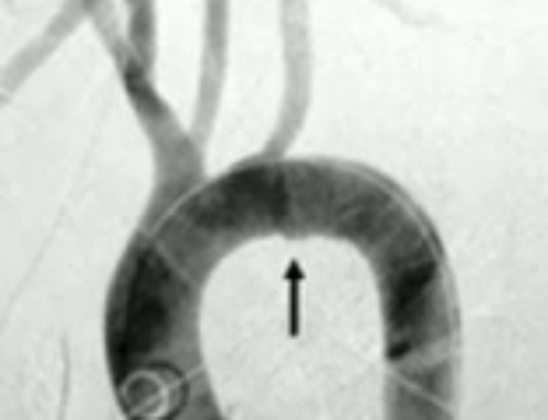
Most common arterial bleeders arise from the anterior division internal iliac artery: superior gluteal, obturator, and internal pudendal artery.
Embolize with whatever you have – coil, PVA, gelfoam. You always prefer to superselect the bleeder and coil it. However, in life threatening, multifocal bleeding, you can opt to coil one or even both internal iliac arteries, although impotence and gluteal pain can be a major side effect.
Gelfoam embolization – since you embolize proximally, you risk non- target embolization such as impotence.
SUMMARY
●Mechanism of injury – Most pelvis injuries require a significant amount of force. The most common mechanisms of injury include motor vehicle collisions and pedestrians struck by a motor vehicle. Associated injuries are frequent with the most common and worrisome being hemorrhage.
●Pelvic fracture types include ring disruptions, sacral fractures, acetabular fractures, and avulsion injuries. Significant hemorrhage may accompany any fracture pattern. Classification schemes are described in the text.
●Physical examination findings associated with an increased risk of pelvic injury include:
•Abnormal position of lower extremities
•Flank, perineal, or scrotal ecchymosis
•Tenderness over the bony pelvis, especially the sacrum and sacroiliac joints
•Focal lower extremity weakness or diminished sensation
•Hematuria or bleeding from the rectum or vagina
Physical examination cannot be relied upon to detect significant pelvis injuries in the patient who is severely injured, intubated, or manifests an altered mental status. Examination of the pelvis to assess stability should be performed gently to avoid displacement of fractures and increased bleeding.
●A bedside ultrasound examination (ie, Focused Assessment with Sonography in Trauma, or FAST) is performed in the great majority of blunt trauma patients. Its role in the assessment of pelvic trauma has yet to be clearly defined.
●Plain radiograph indications – A plain radiograph of the pelvis is obtained in hemodynamically unstable patients; its utility in stable patients and those undergoing computed tomography (CT) is debatable. We do NOT routinely obtain a plain radiograph in patients who meet the following criteria:
•Glasgow coma scale >13
•No pelvic, abdominal, or back complaints
•No tenderness in the lower abdomen, lower back, groin, or bony pelvis
●CT imaging – Multidetector CT scan remains the preferred method for the evaluation of all hemodynamically stable patients with pelvic trauma.
●Stabilization of injured pelvis – Significant pelvis injuries should be immobilized using either a sheet or a commercial pelvic binder wrapped circumferentially around the greater trochanters. The goal is to stabilize injuries; over-reduction of fractures by wrapping too tightly must be avoided.
●Management algorithm – An algorithm for the management of blunt trauma patients with a significant pelvic fracture is provided. Hemodynamically unstable patients with a positive FAST exam are treated with emergency celiotomy, pelvic stabilization, and/or preperitoneal packing; hemodynamically unstable patients with a negative FAST exam can be evaluated with a diagnostic peritoneal aspirate (DPA). If the DPA is positive, the patients go for emergency celiotomy. For patients with a negative FAST and/or DPA, options include pelvic stabilization, preperitoneal packing, or resuscitative endovascular balloon occlusion of the aorta (REBOA). Most patients ultimately undergo pelvic angiography.
●Consultation and transfer – Major pelvic injuries require early consultation with trauma and orthopedic surgery or expeditious transfer to a regional trauma center. Early notification of the operating room and angiography suite staff, including the interventional radiologist, can save valuable time in the hemodynamically unstable patients with a pelvic fracture.
Leave a Reply